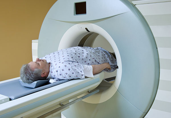
MRI fusion: Ensuring more precise diagnosis and treatment of prostate cancer
June 11, 2024Categories: Blog Posts
 Here’s some good news for men who have an abnormal PSA (prostate-specific antigen) test.
You may not need a biopsy to rule out prostate cancer, thanks to the game-changing use of magnetic resonance imaging (MRI) in diagnosis as well as treatment.
“In the past, we used to biopsy every man who had an abnormal blood test,” says Justin D. Harmon, DO, FACOS, a urologist with Comprehensive Urology Langhorne who was instrumental in developing the MRI fusion prostate biopsy program at St. Mary. “Now, with tools such as MRI and molecular biomarker blood and urine tests, we can determine who really needs a biopsy so we don’t put patients through unnecessary procedures.”
Revolutionizing prostate cancer diagnosis
“The use of MRI has definitely revolutionized our diagnostic ability for prostate cancer,” says Brian Rosenthal, DO, FACOS, also with Comprehensive Urology Langhorne. “If an MRI shows an abnormality, the radiologist gives each lesion a score, based on their level of concern, to determine the need for biopsy. If the lesion is low risk, a biopsy may not be needed.”
If a biopsy is indicated, Harmon and Rosenthal perform the MRI fusion prostate biopsy. This technique fuses pre-biopsy magnetic resonance images of the prostate with ultrasound-guided biopsy images in real time. This gives urologists a clearer view of both normal tissue and of suspicious lesions.
“With MRI fusion, we’re able to target a lesion very specifically, even if it’s the size of a pea,” Harmon notes.
In the past, urologists used a battleship grid of the prostate, targeting 12 samples evenly spaced across the prostate. “It was a blind procedure,” Harmon says. “We couldn’t target anything specific. Now with MRI, we have a highly precise and targeted approach.”
“If you do a targeted fusion biopsy based on MRI findings and it’s negative, you can be pretty confident that you’re not missing the cancer,” Rosenthal notes. “In the past when we were doing random biopsies, you could be missing the cancer. This is definitely a far more precise way to identify prostate cancer.”
St. Mary offers patients more options
If a biopsy is indicated, St. Mary offers patients a choice of two procedures:
- The trans-rectal route. The probe goes into the rectum and the needles traverse the rectal wall.
- The trans-perineal route. The probe goes into the skin under the scrotum, completely avoiding the rectum. “Not all hospitals offer the trans-perineal option, as we do at St. Mary,” Harmon emphasizes. “It has important advantages for the patient, including less rectal bleeding risks and less infection risks the needle does not enter the rectum.”
“We’re very proud of the MRI fusion program that we’ve developed at St. Mary over the past seven years,” Harmon continues. “It’s undergone a tremendous amount of evolution to get to where we are today. We trialed a number of systems to make sure we acquired the best, most precise one. We also did our own internal quality improvement with the aid of our pathologists to make sure we are getting the best results and the best care for our patients.”
Active surveillance for low-grade cancer
It’s important to note that not every man who is diagnosed with prostate cancer needs treatment.
“We often recommend active surveillance for men with low-grade cancer,” Rosenthal says. “This means we follow them with either repeat biopsies down the road or repeat PSA tests, as well as blood and urine biomarkers. This is designed for young men who may be concerned about preserving sexual function and continence, or who don’t yet want to undergo treatment for such a small amount of cancer. It’s also appropriate for older patients who just want to ‘keep an eye on it’ for now. This is a totally reasonable approach.”
If you think you may be at risk for prostate cancer, contact Comprehensive Urology Langhorne for a consultation.
